
Automatic Detection and Segmentation of Crohn’s Disease Tissues From Abdominal MRI
Product Description
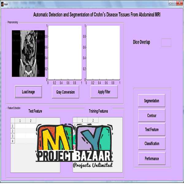
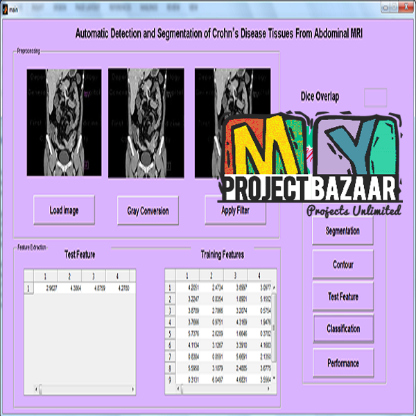
Automatic Detection and Segmentation of Crohn’s Disease Tissues From Abdominal MRI
Abstract—We propose an information processing pipeline for segmenting parts of the bowel in abdominal magnetic resonance images that are affected with Crohn’s disease. Given a magnetic resonance imaging test volume, it is first oversegmented into supervoxels and each supervoxel is analyzed to detect presence of Crohn’s disease using random forest (RF) classifiers.< Final Year Project > The supervoxels identified as containing diseased tissues define the volume of interest (VOI). All voxels within the VOI are further investigated to segment the diseased region. Probability maps are generated for each voxel using a second set of RF classifiers which give the probabilities of each voxel being diseased, normal or background. The negative log-likelihood of these maps are used as penalty costs in a graph cut segmentation framework. Low level features like intensity statistics, texture anisotropy and curvature asymmetry, and high level context features are used at different stages. Smoothness constraints are imposed based on semantic information (importance of each feature to the classification task) derived from the second set of learned RF classifiers. Experimental results show that our method achieves high segmentation accuracy with Dice metric values of 0.90 ± 0.04 and Hausdorff distance of 7.3 ± 0.8 mm. Semantic information and context features are an integral part of our method and are robust to different levels of added noise.
Including Packages
Our Specialization
Support Service
Statistical Report

satisfied customers
3,589
Freelance projects
983
sales on Site
11,021
developers
175+Additional Information
| Domains | |
|---|---|
| Programming Language |
Would you like to submit yours?






There are no reviews yet