Microscopy blood cell segmentation
Product Description
Microscopy blood cell segmentation
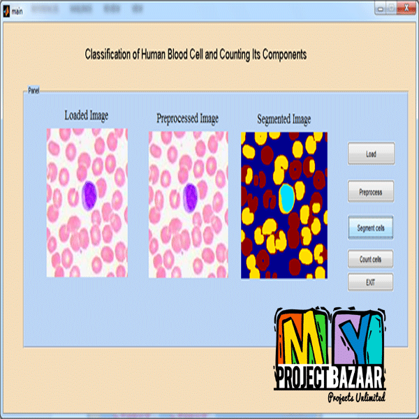
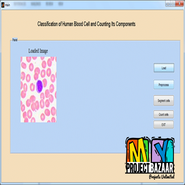
Abstract—Digital Holographic Microscopy (DHM) is becoming recently very popular for cell imaging. The main advantage of digital holographic microscopy over classical microscopy techniques is that it does not only provide the projected image of the object but also provides three dimensional information of the object’s optical thickness. DHM technology could be the core of a label-free imaging for hematology applications.< Final Year Projects > In an ideal framework, a blood sample can be imaged using DHM, machine learning approaches can be used for the cell extraction, differentiation and consequently computing all the relevant blood statistics such as the Mean Corpuscular Volume (MCV), the Red Blood Cell (RBC) count, Red Blood Cell Distribution Width (RDW). The most vital component in such a framework is accurate extraction of the cells. This paper presents a novel approach to cell segmentation in which a probabilistic boosting tree classifier is trained to detect the centers of the cells using Haar-Features. The detected cell centers are used to trigger a marker-controlled power watershed segmentation to compute the cell boundaries. Additionally, we present a comprehensive evaluation of segmentation methods for cell extraction in digital holographic images.
Including Packages
Our Specialization
Support Service
Statistical Report

satisfied customers
3,589
Freelance projects
983
sales on Site
11,021
developers
175+Additional Information
| Domains | |
|---|---|
| Programming Language |

