Automatic Brain Tumor Tissue Detection based on Hierarchical Centroid Shape Descriptor in T1-weighted MR images.
Product Description
Automatic Brain Tumor Tissue Detection based on Hierarchical Centroid Shape Descriptor in T1-weighted MR images
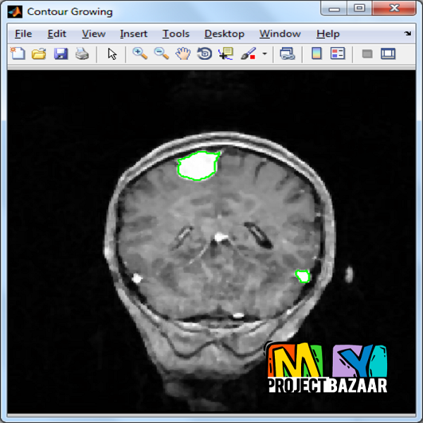
Abstract— The brain tumor tissue detection allows to localize a mass of abnormal cells in a slice of Magnetic Resonance (MR). The automatization of this process is useful for post processing of the extracted region of interest like the tumor segmentation. In order to detect this abnormal growth of tissue in an image, this paper presents a novel scheme which uses a two-step procedure; the k-means method and the Hierarchical Centroid Shape Descriptor (HCSD). The clustering stage is applied to discriminate structures based on pixel intensity while the HCSD allow to select only those having a specific shape. A bounding box is then automatically placed to delineate the region in which the tumor was found. Compared to the tumor delineation performed by an expert, a similarity measure of 91% was reached by using the Dice coefficient. The tests were carried out on 254 T1-weighted MRI images of 14 patients with brain tumors. It offers the advantage to be a noninvasive technique that enables the analysis of brain tissues. The early detection of tumor in the brain leads on saving the patients’ life through proper care. Due to the increasing of medical data flow, the accurate detection of tumors in the MRI slices becomes a fastidious task to perform. Furthermore the tumor detection in an image is useful not only for medical experts, but also for other purposes like segmentation and 3D reconstruction. The method proposed in this work allows to automatically and accurately detect the abnormal tissues in preoperative images. The man-ual delineation and visual inspection will be limited in order to avoid time consumption by medical doctors. < final year projects >
Including Packages
Our Specialization
Support Service
Statistical Report

satisfied customers
3,589
Freelance projects
983
sales on Site
11,021
developers
175+Additional Information
| Domains | |
|---|---|
| Programming Language |

