Classification of Color Images of Dermatological Ulcers
Product Description
Classification of Color Images of Dermatological Ulcers
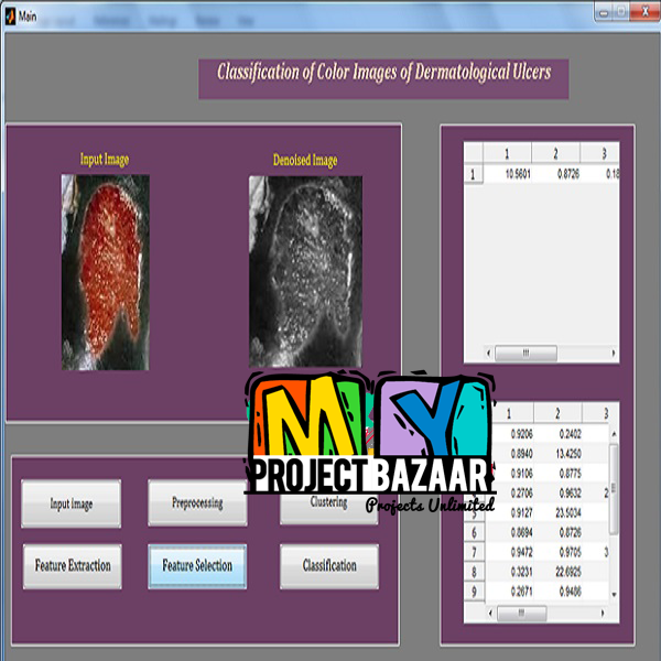
Abstract—We present color image processing methods for the analysis of images of dermatological lesions. The focus of this study is on the application of feature extraction and selection methods for classification and analysis of the tissue composition of skin lesions or ulcers, in terms of granulation (red), fibrin (yellow), necrotic (black), callous (white), and mixed tissue composition.< Final Year Project >The images were analyzed and classified by an expert dermatologist into the classes mentioned previously. Indexing of the images was performed based on statistical texture features derived from cooccurrence matrices of the red, green, and blue (RGB), hue, saturation, and intensity (HSI), L*a*b*, and L*u*v* color components. Feature selection methods were applied using the Wrapper algorithm with different classifiers. The performance of classification was measured in terms of the percentage of correctly classified images and the area under the receiver operating characteristic curve, with values of up to 73.8% and 0.82, respectively.
Including Packages
Our Specialization
Support Service
Statistical Report

satisfied customers
3,589
Freelance projects
983
sales on Site
11,021
developers
175+Additional Information
| Domains | |
|---|---|
| Programming Language |
Would you like to submit yours?


There are no reviews yet